Draft of chapter 6, Spike initiation with an initial segment

I have just uploaded an incomplete draft of chapter 6, "Spike initiation with an initial segment". This chapter deals with how spikes are initiated in most vertebrate neurons (and also some invertebrate neurons), where there is a hotspot of excitability close to a large soma. This situation has a number of interesting implications which make spike initiation quite different from the situation investigated by Hodgkin and Huxley, that of stimulating the middle of an axon. Most of the chapter describes the theory that I have developed to analyze this situation, called "resistive coupling theory" because the axonal hotspot is resistively coupled to the soma.

The chapter is currently unfinished, because a few points require a little more research, which we have not finished. The presentation is also a bit more technical than I would like, so this is really a draft. I wanted nonetheless to release it now, as I have not uploaded a chapter for a while and it could be some time before the chapter is finished.

Technical draft for chapter 5, Propagation of action potentials

I have just uploaded a technical draft on chapter 5 of my book on action potentials: Propagation of action potentials. This draft introduces the cable equation, and how conduction velocity depends on axon diameter in unmyelinated and myelinated axons. There is also a short section on the extracellular potential. There are a few topics I want to add, including branching and determinants of conduction velocity (beyond diameter). There is also (almost) no figure at the moment. Finally, it is likely that the chapter is reorganized for clarity. I wanted to upload this chapter nonetheless so as to move on to the next chapter, on spike initiation with an initial segment.

New chapter : Excitability of an isopotential membrane

I have just uploaded a new chapter of my book on the theory of action potentials: Excitability of an isopotential membrane. In this chapter, I look mostly at the concept of spike threshold: the different ways to define it, its quantitative relation to different biophysical parameters (eg properties of sodium channels), and the conditions for its existence (eg a sufficient number of channels). This is closely related to my previous work on the threshold equation (Platkiewicz and Brette, 2010). It also contains some unpublished work (in particular updates of the threshold equation).

I am planning to extend this chapter with:

  • A few Brian notebooks.
  • A section on excitability types (Hodgkin classification).
  • Some experimental confirmations of the threshold equation that are under way (you will see in section 4.4.2 that current published experimental data do not allow precise testing of the theory).

I am now planning to work on the chapter on action potential propagation.

All comments are welcome.

Update on the book

I am writing a book on the theory of action potentials. As I haven't published any new chapter for about 6 months, I will give a brief update on the progress of the book.

It turns out that writing a book is difficult (!). As I write the book, I get to question some aspects I take for granted and I realize they are actually not that simple. That I have learned a lot about biophysics. In turn, this tends to make the book more complicated, and so it requires some additional digestion work. I also realize that some important questions are just unresolved and require some research work. Finally, it is quite difficult to write a book while continuing the normal course of research. I started with a one hour per day habit, but this may not be optimal; I tend to spend the first half-hour trying to get back to the matter of the book. I am starting a new routine with two mornings twice a week. We will see how it goes!

These last months I have been working on the 4th chapter, on excitability of an isopotential membrane. This will contain in particular some of the material in Platkiewicz & Brette (2010) on the relation between biophysical parameters and threshold. I wanted to use the same theoretical approach to apply it to other aspects of the action potential (speed, peak etc), so that needed some more work. I realized that some approximations we had done could be enhanced, but the math is slightly more complicated. It is a challenge to keep this chapter simple. I also wanted to apply the theory to the original Hodgkin-Huxley model, but unfortunately it works moderately well. One reason is that the model was fitted to the full action potential and not to initiation (as in fact essentially all empirical neuron models). So in particular, H&H experiments show that the Na+ conductance depends exponentially on voltage at low voltage, but their model doesn't (or approximately does with a different factor). Another reason is the K+ channel has less delay than in the actual squid axon (which they acknowledge), so there is some interaction with the initial Na+ current. So I will go with a simpler approach, using a simplified excitability model. Neurons are not isopotential anyway.

I am also planning to reorganize and partly rewrite chapters 2 and 3. I find the current chapter 3 (action potential of an isopotential membrane) a bit too technical. I also want to change chapter 2 (membrane polarization) to talk about alternative theories of the membrane potential (Tasaki, Ling, etc). Then I want to insert a chapter on the ionic channel theory of action potentials, which explains the theory and discusses empirical evidence, before the chapter on the Hodgkin-Huxley model. Generally, I want to simplify the exposition. But given my rather slow progress on the book so far, I will probably postpone this and first write drafts of the following chapters.

Finally, I have worked a bit on energy consumption and pumps, and I found out that the current treatment in the literature is not entirely satisfactory (see for example my comments on a classical paper on the subject). It turns out that it is a pretty complicated problem (especially systems of pumps).

In brief, I am trying to finish a first version of the chapter on excitability of an isopotential membrane, hopefully within one month.

Comments on the soliton model of action potential

Recently a radically new biophysical theory of action potentials has been proposed, which I will call here the “soliton theory”, according to which action potential propagation is a travelling pulse (soliton) of membrane (lipidic) lateral density, i.e., a sound wave along the axon membrane (Heimburg and Jackson, 2005; Andersen et al., 2009). In this theory, the lipid membrane undergoes a phase transition, from liquid to gel, which propagates along the axon. The electrical spike is then attributed to piezoelectric effects (mechanical changes inducing electrical potential changes). No role is attributed to proteins (ionic channels).

I start with some positive comments. Usually only the electrochemical aspects of neural excitability are considered in the field. But it is known that the electrical phenomenon is accompanied by mechanical, optical and thermal effects, which are the main focus of the soliton theory. The theory also has the merit of bringing attention to non-electrical phenomena in biological membranes, such as structural changes and mechanical effects, which are typically ignored in the field.

The theory appears to be motivated by the observation of reversible heat release during the action potential, i.e., heat is released in the rising phase of the spike and absorbed in the falling phase. Quantitatively, release and absorption have the same magnitude (experimental precision being in the range of 10-20% according to the original papers). This is not explained by HH theory. It does not mean, however, that it is contradictory with HH theory; rather, it would require some additional mechanism that is not part of the theory (there are some speculations in the literature, by Hodgkin, Keynes, Tasaki, and probably others). HH theory does not directly address mechanical changes accompanying the spike, but it does imply mechanical changes by at least two plausible mechanisms: 1) osmotic effects, i.e., water enters the cell along with Na+ influx, and exist with K+ outflux, leading to a diameter variation in phase with the spike (see e.g. (Kim et al., 2007); 2) an electrical field applied on the membrane can change the curvature of the membrane. The appeal of the soliton theory is that a sound wave produces reversible heat and mechanical variations; electrical variation is attributed to piezoelectric effects and therefore all these effects should be in phase. Thus in a way, it has some theoretical elegance. Of course, the actual biophysical mechanisms are not necessary elegant.

Let us now examine the premises and predictions of the theory. First, it is assumed that the lipidic membrane is close to a melting transition, which the authors claim occurs slightly below the body temperature of 37°C. It is rather surprising to read this starting point when the theory is meant to address the shortcomings of the HH model. Let us recall that the HH model is a model of the giant axon of squid, which is a cold-blooded animal living in the ocean. Body temperature is thus much colder and variable. But this is not the most problematic aspect of the theory; let us assume for now that the squid membrane does have the required property.

The main quantitative prediction of the theory is conduction velocity, which follows from membrane properties, and it is calculated to be around 100 m/s. The conclusion is that there is “a minimum velocity of the solitons that is close to the propagation velocity in myelinated nerves”. First, the squid axon is not myelinated (it is anyway not clear why the theory should apply to myelinated nerves rather than unmyelinated ones), and conduction velocity is around 20 m/s. In any case, 100 m/s is not the propagation velocity in myelinated nerves. It is the upper bound of conduction velocity in nerve, which varies over several orders of magnitude and is much smaller for most axons. It actually varies with diameter, quite in line with predictions from HH and cable theory (scaling with square root of diameter for unmyelinated axons; with diameter for myelinated axons). One of the main predictions of the 1952 HH paper where the model is described is conduction velocity, which is accurate within 20% (Hodgkin and Huxley, 1952); to be compared with 500% error in the soliton theory. The prediction was calculated as follows (see chapter 3 of my book in progress): the HH model was built and fitted on a space-clamped (isopotential) squid axon; then it was extended to a model of propagating spike with the cable formalism, ie by adding the axial current term (based on measurement of diameter and intracellular resistivity); then the model was run and a conduction velocity of around 20 m/s was found.

If one of the main predictions of the soliton theory is a minimum conduction velocity of around 100 m/s, then it is definitely wrong. There are of course many other aspects of the theory that are very problematic. HH theory is essentially the ionic hypothesis, ie the idea that changes in membrane potential are due to ionic and capacitive transmembrane currents. There have been numerous quantitative tests of this hypothesis, such as: the peak of the spike is well predicted by the Nernst potential of Na+, the influx of Na+ and outflux of K+ per spike (measured with radioactive tracers) are predicted by the HH model; fluorescence imaging now shows influxes of Na+ in phase with spikes. Early work by HH and colleagues have showed that the squid axon is inexcitable when extracellular Na+ is replaced by choline. All this body of work, which includes many accurate quantitative predictions of HH theory, is contradictory with the soliton theory. The authors seem to deny the existence of ionic channels, which is extremely strange. The detailed molecular and genetic structure of ionic channels is known, as well as their electrophysiological properties (see e.g. (Hille, 2001)); drugs targetting Na+ channels (for which there is huge empirical evidence) block action potentials. There is also the Na/K pump, a major contributor of energy consumption in neurons, which maintains the Na+/K+ concentration gradients necessary in the ionic hypothesis, and which seems totally absurd in the soliton theory.

As it stands, the soliton theory of spikes is contradictory with an extremely large body of experimental evidence, which is explained by HH theory (ie the ionic hypothesis) (note that there have been a number of alternative theories, eg by Ling and Tasaki).


Update (21.7.2016): A great resource about the evidence in favor of the ionic hypothesis is (Hodgkin, 1951). There is in fact a section dealing with heat production, where it is by the way noted that overall nerve activity does produce heat, but a quite small amount. It is also clear in the text that heat production in the ionic hypothesis is not that of the equivalent electrical circuit that is used to present the HH model – which is only equivalent in terms of the mathematical equations describing the currents, not physically. That is, the axial current does follow the expectation from the electrical circuit, since in the theory it is due to the electrical field in an electrolyte, but not the transmembrane current, which corresponds to mixing of extra- and intra-cellular solutions in addition to field effects (plus the at the time unknown mechanisms of permeability changes).


Andersen SSL, Jackson AD, Heimburg T (2009) Towards a thermodynamic theory of nerve pulse propagation. Prog Neurobiol 88:104–113.

Heimburg T, Jackson AD (2005) On soliton propagation in biomembranes and nerves. Proc Natl Acad Sci U S A 102:9790–9795.

Hille B (2001) Ion Channels of Excitable Membranes. Sinauer Associates.

Hodgkin A, Huxley A (1952) A quantitative description of membrane current and its application to conduction and excitation in nerve. J Physiol Lond 117:500.

Hodgkin AL (1951) The Ionic Basis of Electrical Activity in Nerve and Muscle. Biol Rev 26:339–409.

Kim GH, Kosterin P, Obaid AL, Salzberg BM (2007) A mechanical spike accompanies the action potential in Mammalian nerve terminals. Biophys J 92:3122–3129.

General bibliography on action potential theory

Two general introductory biology textbooks, covering the excitability of neurons and muscles are (Matthews, 2002) and (Keynes et al., 2011). The biophysics and modeling of neurons are covered in (Johnston and Wu, 1994) and (Sterratt et al., 2011). Both are quite accessible and include all essential material including compartmental modeling of dendrites.

There is a great book that covers many topics in cell biology from a physicist perspective, including excitability: Physical biology of the cell (Phillips et al., 2008).

There are two excellent reviews by Hodgkin that are particularly useful to understand the experimental basis of Hodgkin-Huxley theory, including myelinated axons: (Hodgkin, 1951, 1964).

The reference textbook for the biophysics ionic channels is (Hille, 2001). (Johnston and Wu, 1994) also includes some material about stochastic analysis of channels.

Linear cable theory is covered in great detail in (Tuckwell, 1988) and (Koch, 1999). An excellent review by one of the historical figures of cable theory is (Rall, 2011). (Jack et al., 1975) also covers cable theory including active axonal conduction, and it also includes muscle APs and their propagation and classic theory of excitability (threshold).

Theory of electro-osmosis (interaction between osmosis and electrical field) is treated in (Hoppensteadt and Peskin, 2004).


Hille B (2001) Ion Channels of Excitable Membranes. Sinauer Associates.

Hodgkin AL (1951) The Ionic Basis of Electrical Activity in Nerve and Muscle. Biological Reviews 26:339–409.

Hodgkin AL (1964) The conduction of the nervous impulse. C. C. Thomas.

Hoppensteadt FC, Peskin C (2004) Modeling and Simulation in Medicine and the Life Sciences, 2nd edition. New York: Springer.

Jack JB, Noble D, Tsien R (1975) Electric Current Flow in Excitable Cells. Oxford: OUP Australia and New Zealand.

Johnston D, Wu SM-S (1994) Foundations of Cellular Neurophysiology, 1 edition. Cambridge, Mass: A Bradford Book.

Keynes RD, Aidley DJ, Huang CL-H (2011) Nerve and Muscle, 4 edition. Cambridge ; New York: Cambridge University Press.

Koch C (1999) Biophysics of computation: Information processing in single neurons. Oxford University Press, USA.

Matthews GG (2002) Cellular Physiology of Nerve and Muscle, 4 edition. Osney Mead, Oxford ; Malden, MA: Wiley-Blackwell.

Phillips R, Kondev J, Theriot J (2008) Physical Biology of the Cell, 1 edition. New York: Garland Science.

Rall W (2011) Core Conductor Theory and Cable Properties of Neurons. In: Comprehensive Physiology. John Wiley & Sons, Inc.

Sterratt D, Graham B, Gillies DA, Willshaw D (2011) Principles of Computational Modelling in Neuroscience, 1 edition. Cambridge; New York: Cambridge University Press.

Tuckwell H (1988) Introduction to theoretical neurobiology, vol 1: linear cable theory and dendritic structure. Cambridge: Cambridge University Press.


A book on the theory of action potentials

Latest news

I am writing a book on the theory of action potentials. I will post chapters on this website as I write them. Note however that this is essentially preparatory work for the book, and I am probably going to rewrite and reorganize it quite extensively. So do not expect a very well organized and didactical text at this stage; only the essential content should remain. I would be happy to read your comments, in particular if you find errors or omissions: please let me know and I will reward you with your name in the acknowledgments!

The plan is to start with standard biophysics of excitability, and then I will expose more advanced topics, such as: how spikes initiate in real life (as opposed to when you stimulate an axon), how excitability changes on different time scales, and how a cell learns to spike. The book adopts a systemic viewpoint; that is, the goal is to understand how the coordination of channels creates and maintains functional action potentials. I would also like to give an epistemological flavor to it that I find is missing in most textbooks: what is a model, how is it built and tested, what is its empirical value, etc.

Why this book and who is it for? With this book, I am hoping to bring theoreticians to the field of neural excitability, and to give them the necessary material that is currently scattered over many references. Currently, the field is largely dominated by experimental biologists. Yet, as I will try to convey, this is a field where one can ask many key neuroscientific questions in a context where the link between structure and function is much less speculative than in neural network research, including questions of learning and adaptation, and where one can actually develop quantitative, testable theories. As a bonus, I would also be happy if I could manage to convey some elements of theory to biologists.

Prerequisites. In principle you do not need to know much about biology to read this book, as I will try to introduce the necessary information. I am expecting some mathematical skills, mostly calculus and basics of differential equations, but nothing very advanced. Regarding physics, electrophysiology is obviously a lot about electricity. In the current version, I am assuming the reader has some basic knowledge of electricity (what current and charges are, Ohm's law). But I am planning to add a primer on electricity.

Each chapter is accompanied by a set of examples using the Brian 2.0 simulator, in the form of Jupyter notebooks.

I am also compiling a general bibliography on action potential theory (books and reviews only).

Here is a tentative outline (available chapters are in bold):

  1. Action potentials. An overview of action potentials, their scientific history and their function. Brian notebooks for chapter 1Last update: 9.6.2016
  2. The membrane potential. The biophysical basis of membrane polarization. Brian notebooks for chapter 2Last update: 9.6.2016
  3. Action potential of an isopotential membrane. Basic biophysics of the squid giant axon and Paramecium, and the Hodgkin-Huxley model. Brian notebooks for chapter 3Last update: 19.7.2016
  4. Excitability of an isopotential membrane. Theoretical analysis of excitability in isopotential models [Some content to be added on excitability types]. Last update: 14.3.2017.
  5. Propagation of action potentials (technical draft). The cable equation; active propagation in unmyelinated and myelinated axons. Last update: 13.4.2017.
  6. Spike initiation in the axonal initial segment (incomplete draft). Excitation through the soma and AIS, as opposed to excitation of the middle of a squid axon. Last update: 5.4.2018.
  7. Dynamics of excitability. How excitability (spike threshold) changes at different time scales (adaptation and plasticity).
  8. Energy consumption.
  9. Learning to spike. How a cell builds and maintains a functional spiking system.


Why do neurons spike?

Why do neurons produce those all-or-none electrical events named action potentials?

One theory, based on the coding paradigm, is that the production of action potentials is like analog-to-digital conversion, which is necessary if a cell wants to communicate to a distant cell. It would not be necessary if neurons were only communicating with their neighbors. For example, in the retina, most neurons do not spike but interact through graded potentials, and only retinal ganglion cells produce spikes, which travel over long distances (note that there is actually some evidence of spikes in bipolar cells). In converting graded signals into discrete events, some information is lost, but that is the price to pay in order to transmit any signal at all over a long distance. There is some theoretical work on this trade-off by Manwani and Koch (1999).

Incidentally, this theory is sometimes (wrongly) used to argue that spike timing does not matter because spikes are only used as a proxy for an analog signal, which is reflected by the firing rate. This theory is probably not correct, or at least incomplete.

First, neurons start spiking before they make any synaptic contact, and that activity is important for normal development (Pineda and Ribera, 2009). Apparently, normal morphology and mature properties of ionic channels depend on the production of spikes. In many neuron types, those early spikes are long calcium spikes.

A more convincing argument to me is the fact that a number of unicellular organisms produce spikes. For example, in paramecium, calcium spikes are triggered in response to various sensory stimuli and trigger an avoidance reaction, where the cell swims backward (reverting the beating direction of cilia). An interesting point here is that those sensory stimuli produce graded depolarizations in the cell, so from a pure coding perspective, the conversion of that signal to an all-or-none spike in the same cell seems very weird, since it reduces information about the stimuli. Clearly, coding is the wrong perspective here (as I have tried to argue in my recent review on the spike vs. rate debate). The spike should not be seen as a code for the stimulus, but rather as a decision or action, in this case to reverse the beating direction. This argues for another theory, that action potentials mediate decisions, which are by definition all-or-none.

Action potentials are also found in plants. For example, mimosa pudica produces spikes in response to various stimuli, for example if it is touched, and those spikes mediate an avoidance reaction where the leaves fold. Those are long spikes, mostly mediated by chloride (which is outward instead of inward). Again the spike mediates a timed action. It also propagates along the plant. Here spike propagation allows organism-wide coordination of responses.

It is also interesting to take an evolutionary perspective. I have read two related propositions that I found quite interesting (and neither is about coding). Andrew Goldsworthy proposed that spikes started as an aid to repair a damaged membrane. There is a lot of calcium in the extracellular space, and so when the membrane is ruptured, calcium ions rush into the cell, and they are toxic. Goldsworthy argues that the flow of ions can be reduced by depolarizing the cell, while repair takes place. We can immediately make two objections: 1) if depolarization is mediated by calcium then this obviously has little interest; 2) to stop calcium ions from flowing in, one needs to raise the potential to the reversal potential of calcium, which is very high (above 100 mV). I can think of two possible solutions. One is to trigger a sodium spike, but it doesn't really solve problem #2. Another might be to consider evenly distributed calcium channels on the membrane, perhaps together with calcium buffers/stores near them. When the membrane is ruptured, lots of calcium ions enter through the hole, and the concentration increases locally by a large amount, which probably immediately starts damaging the cell and invading it. But if the depolarization quickly triggers the opening of calcium channels all over the membrane, then the membrane potential would increase quickly with relatively small changes in concentration, distributed over the membrane. The electrical field then reduces the ion flow through the hole. It's an idea, but I'm not sure the mechanism would be so efficient in protecting the cell.

Another related idea was proposed in a recent review. When the cell is ruptured, cellular events are triggered to repair the membrane. Brunet and Arendt propose that calcium channels sensitive to stretch have evolved to anticipate damage: when the membrane is stretched, calcium enters through the channels to trigger the repair mechanisms before the damage actually happens. In this theory, it is the high toxicity of calcium that makes it a universal cellular signal. The theory doesn't directly explain why the response should be all-or-none, however. An important aspect, maybe, is cell-wide coordination: the opening of local channels must trigger a strong enough depolarization so as to make other calcium channels open all over the membrane of the cell (or at least around the stretched point). If the stretch is very local, then this requires an active amplification of the signal, which at a distance is only electrical. In other words, fast coordination at the cell-wide level requires a positive electrical feedback, aka an action potential. Channels must also close (inactivate) once the cellular response has taken place, since calcium ions are toxic.

Why would there be sodium channels? It's actually obvious: sodium ions are not as toxic as calcium and therefore it is advantageous to use sodium rather than calcium. However, this is not an entirely convincing response since in the end, calcium is in the intracellular signal. But a possible theory is the following: sodium channels appear whenever amplification is necessary but no cellular response is required at that cellular location. In other words, sodium channels are useful for quickly propagating signals across the cell. It is interesting to note that developing neurons generally produce calcium spikes, which are then converted to sodium spikes when the neurons start to grow axons and make synaptic contacts.

These ideas lead us to the following view: the primary function of action potentials is cell-wide coordination of timed cellular decisions, which is more general than fast intercellular communication.

Why the textbook account of spike initiation is wrong

In previous posts, I have tried to explain why spike initiation is “sharp” or abrupt, basically as in an integrate-and-fire model. The explanation relies on the fact that spikes are initiated in the axon, next to the soma. What may not be so obvious is how this differs from the textbook account of spike initiation. That textbook account would go as follows: first, the spike is initiated at a distal site in the axon, through the interplay between transmembrane currents, then the spike is propagated to the soma. On the course of its propagation, its shape changes and becomes sharper (see the explanation in this paper for example).

This account is wrong on several levels. But fundamentally, it has to do with the inadequacy of the transportation metaphor. Let us take a step back. What do we mean when we say that a spike propagates along an axon? In fact, not much actually moves physically along the axon. There is very little ion movement in the longitudinal direction. Ions move mostly through the membrane, in the radial direction. It produces a spatiotemporal pattern of electrical activity, and the reason we say a spike propagates is that in certain conditions that pattern is a solitary wave for which we can define a velocity. This is actually how Hodgkin and Huxley predicted spike velocity in the squid giant axon, by postulating that the potential V(x,t) can be written as U(x-v.t), and then looking for the velocity v that was consistent with their equations. This velocity has actually nothing to do with the velocity of the ions that carry the electrical charges. So basically we often describe electrical activity in axons in terms of transportation of spikes, but one should keep in mind that this is just a metaphor.

Now when we say that there is a spike that moves and gets transformed on its way, we should realize that the metaphor has reached its limits. As nothing actually gets transported, we are not saying anything else than there is an irregular spatiotemporal pattern of activity. That is, we are not saying anything at all. That is all the more true when we are talking of time scales shorter than the duration of a spike (as in the backprogation from initiation site to soma).

The transportation metaphor, unfortunately, is highly misleading. It leads to incorrect reasoning in this case. Here is the reasoning in question. The onset of a spike at the axonal initiation site is smooth. The onset of a spike at the soma is sharp. Therefore, the spike onset gets sharper through the propagation from initiation site to soma. On the surface, the reasoning seems flawless. But now here is a disturbing fact: from the spike shape at the axonal initiation site, I can make a quantitative prediction of onset rapidness at the soma, without knowing anything of what's in between (that's in a paper being submitted). Therefore, onset rapidness at the soma is determined by properties of spike initiation, not of propagation. How can that be?

The flaw is this: in the transportation metaphor, somatic and axonal spikes are implicitly seen as local events both in space and time, which can then be related by the transportation metaphor (object at place A and time t1 travels to place B at time t2). As mentioned above, the relevance of the transportation metaphor is questionable at this spatial and temporal scale, all the more when what is being transported changes. What I showed in my previous paper and review is precisely that spike initiation is not local. It is determined both by the properties of local Na channels and by resistive properties of the coupling between soma and axonal initiation site, which are not local. For example, if you moved the Na channels away from the soma, the neuron would become more excitable, even though local properties at the initiation site would be identical. Spike initiation is not a local phenomenon because of the proximity of the axonal initial segment to the big somatodendritic compartment.

Thus the sharpness of somatic spikes is actually determined not by propagation properties, contrary to what was claimed before, but by spike initiation properties. The catch is that those properties are not local but rather encompass the whole soma-initial segment system.

I had previously called this biophysical explanation the “compartmentalization hypothesis”. I realize now that this can be very misleading because physiologists tend to think in terms of initiation in one compartment and transportation from one compartment to the other. I will now use a different terminology: “critical resistive coupling”, which emphasizes that spike initiation relies on a system of coupled compartments.

Sharpness of spike initiation explained with photos of moutain roads

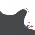
In a previous post, I tried to explain the idea that spike initiation is “sharp” using a few drawings. The drawings represent a ball on a hilly landscape. The position of the ball represents the state of the system (neuron), and the altitude represents its energy. The ball tends to reside in positions of local minima of the energy. Spiking occurs when some energy is added to the system (stimulation with electrode or synaptic input) so that the ball is moved past a hill and then falls down:

Sharp-initiation-2

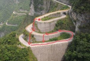
What is not seen in this drawing is what happens next. The membrane potential is reset and the neuron can fire again, in principle, but in the drawing the ball ends up on a lower valley than at the start. Biophysically, that corresponds to the fact part of the energy that was stored in electrochemical gradients of ion concentration across the membrane has been released by the spike, and so indeed the energy is now lower. Spiking again would mean falling down to an even lower valley. But since energy is finite, this has to stop at some point. In reality it doesn't stop because the neuron slowly rebuilds its electrochemical energy by moving ions against their concentration gradients with the sodium/potassium pump, which requires some external energy (provided by ATP). In the drawing, this would correspond to slowly moving up from the low valley to the high valley. Unfortunately you can't represent that in two dimensions, you need to represent a road spiraling up around a hill. This is the best photographic illustration I have found:

Tianmen-road

This is a road in China (Tianmen mountain). The neuron starts on top (red ball). With some external energy, the ball can be moved up past the barrier and then it falls. It lands on the road, but on a lower level. It can be made to spike again. Slowly, continuously, some energy is added to the neuron to rebuild its electrochemical gradients: the ball moves up the road. As long as spikes are rare enough, the neuron can continue to spike at any time (ideally, the road should spiral around the hill but it's close enough!).

Smooth spike initiation would correspond to something like that (a road in the Alps):

Alps-road